Feather Bioaerosol
© 2024 Jeffrey C. May – Small keratin granules on feather fragments may cause allergy or coughing.
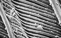 Low-power SEM (500x) of a feather from a cockatiel bird. The granular appearance of the surface of the feather structures is due to bacteria-like organisms, visible below in the higher-power SEM.
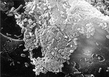
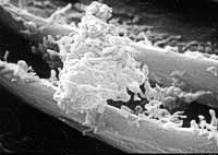 The large triangular shape at the center of this high-power SEM (4000x) is either a bird dander particle or a human skin scale. It is completely covered with keratin granules. Bean-shaped individual organisms are visible all around the dander.
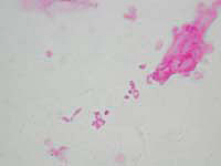 This is a light photomicrograph at 1000x of the particulates that came out of a homeowner’s down pillow. (The particles are stained pink with acid fuchsin.) I believe that these particulates are the same keratin granules found on the bird’s wing. The larger, oblong object is probably bird dander. I believe that inhaling these respirable particles from down pillows and quilts can cause allergy and asthma symptoms.
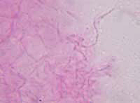
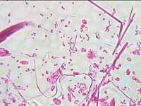 This is a light photomicrograph at 400x of the particulates that came out of a homeowner’s down quilt. (The particles are stained pink with acid fuchsin.) Keratin granules are visible as well as feather fragments. Some of these feather fragments are inhalable and, I believe, can cause coughing.
Bacteria from a Smelly Sponge
© 2024 Jeffrey C. May – The surface of a kitchen sponge that reeked.
 This is a high-power SEM (4000x) from the surface of a kitchen sponge that reeked. Numerous rod-shaped bacteria are visible. Most of the bacteria are embedded in a plane that is draped over the surface of the sponge. Other bacteria that were living in the sponge were deriving their nutrients from milk that had been soaked up by the sponge, but these bacteria are all within the surface of a human skin scale.
In any indoor air sample, the most common aerosol particle is the human skin scale. These are usually well defined, and about 25 microns in size. When viewed through a light microscope after staining, they appear to be pink. Very often in indoor air samples, bacteria can be seen in indistinct pink-stained fragments much smaller than 25 microns. I believe that these are skin-scale fragments that have been partially digested by bacteria in damp reservoirs where dust has accumulated. These reservoirs can include humidifiers, air-conditioners, and carpets or rugs that have been wet.
Humidifiers
© 2024 Jeffrey C. May – A white film on the surface of water in a humidifier.
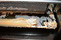 The water tray of the furnace humidifier at the left has a crust of crystalline minerals at its lip. The minerals were deposited from the many gallons of water evaporated by the rotating sponge coated with a honeycomb plastic in the upper part of the picture. The white film floating on the surface of the dark water consists of actinomycetes (bacteria-like organisms).
 At the left is a light photomicrograph (1000 x) of the actinomycetes taken from the surface of the humidifier water. The actinomycetes are stained pink with acid fuchsin. These organisms are normally found growing in soil, and are sometimes responsible for the smell of freshly turned earth. I often find actinomycetes in humidifiers, both on furnaces and in portable room units. Inhalation of the organisms can cause allergy problems and hypersensitivity pneumonitis. Actinomycetes grow as fungi, producing spores and hyphae, though much smaller.
Mites
© 2024 Jeffrey C. May – A mold-eating mite.
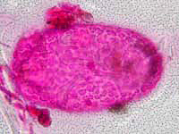 There are hundreds of species of mites. Most dwell in the soil, but some live in our basements and bedrooms. At the left is a light photomicrograph of a 112-micron egg, containing a fully developed mold-eating mite. The entire egg is stained pink by acid fuchsin, but you can see the textured pattern of the egg surface and the mite’s folded legs. There are a few pink-stained mold spores at the left. The egg was found in a colony of mildew growing on a wall in a house.
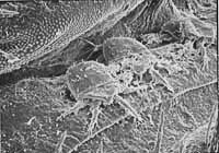 Some mites live only on other organisms, and some forage for their nutrients. Dust mites eat skin scales that we humans shed in abundance and leave in our beds and couches. At the left is a 150x scanning electron micrograph of three mites within a gap between two segments of a dermestid beetle larva. The larva was feeding on the carcass of a dead squirrel.
Stachybotrys Mold
© 2024 Jeffrey C. May – Often referred to as the “toxic black mold.”
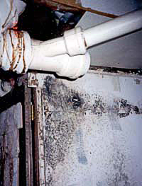 Stachybotrys chartarum is often referred to as “toxic black mold,” although only about a third of the strains actually produce toxins while growing. The black patch on the drywall at the left is Stachybotrys mold growing under a pipe leak. This black mold only grows on cellulose (paper, wood, etc.) that has remained soaked for days, and never grows on non-porous surfaces such as metal or glass, where you can sometimes see other types of black mold growing on dust.
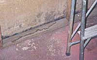 In the photograph at the left, Stachybotrys mold is growing on the drywall just above a two-by-four resting on a concrete basement floor. The stains on the drywall, wood, and floor suggest water leaks. Stachybotrys spores grow in large clumps and adhere to one another within the clump. For this reason, it is relatively rare to find Stachybotrys spores in the air, unless moldy surfaces have been disturbed.